
Plain film x-ray is the most common diagnostic radiological modality used in hospitals today. The axillary view is a substitution for the scapular Y view.
X Rays Joseph J Schreiber Md

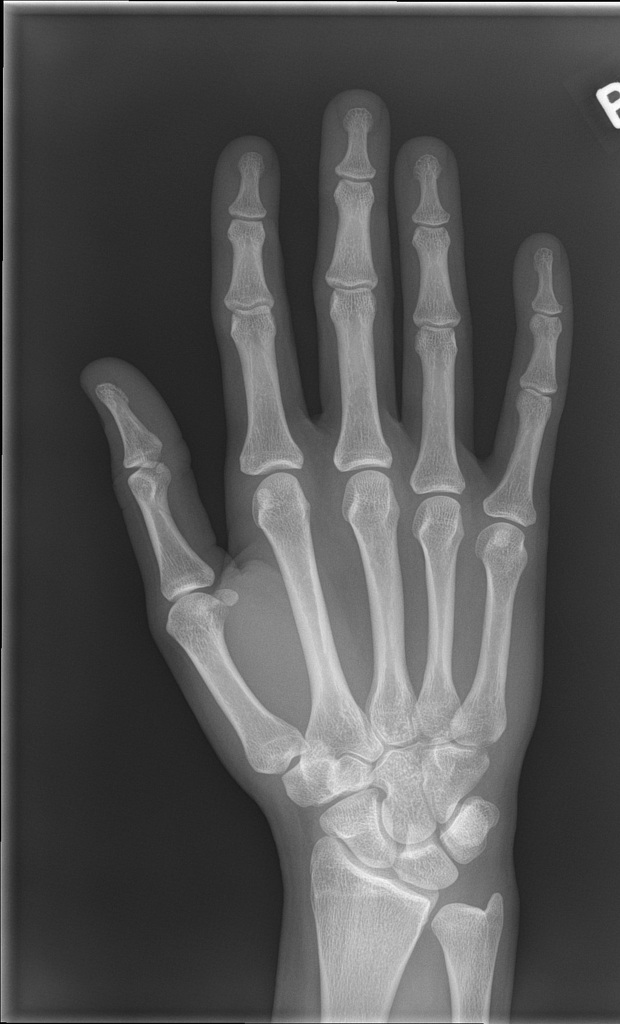
Normal Hand Of 13 Year Old X Ray Stock Image C039 3319 Science Photo Library
Hand X Ray
A faint rounded density is seen in the base of the left lower hemithorax probably representing a nipple shadow.

Normal hand x ray. Normal elbow X-ray - 10 year old. The lungs are well aerated. Click image to align with top of page.
An X-ray is a common imaging test that has been used for decades to help doctors view the inside of the body without having to open it up using surgery. Even if the finger looks normal and can move it may require treatment. You and your hand specialist will determine the best approach for your individual situation.
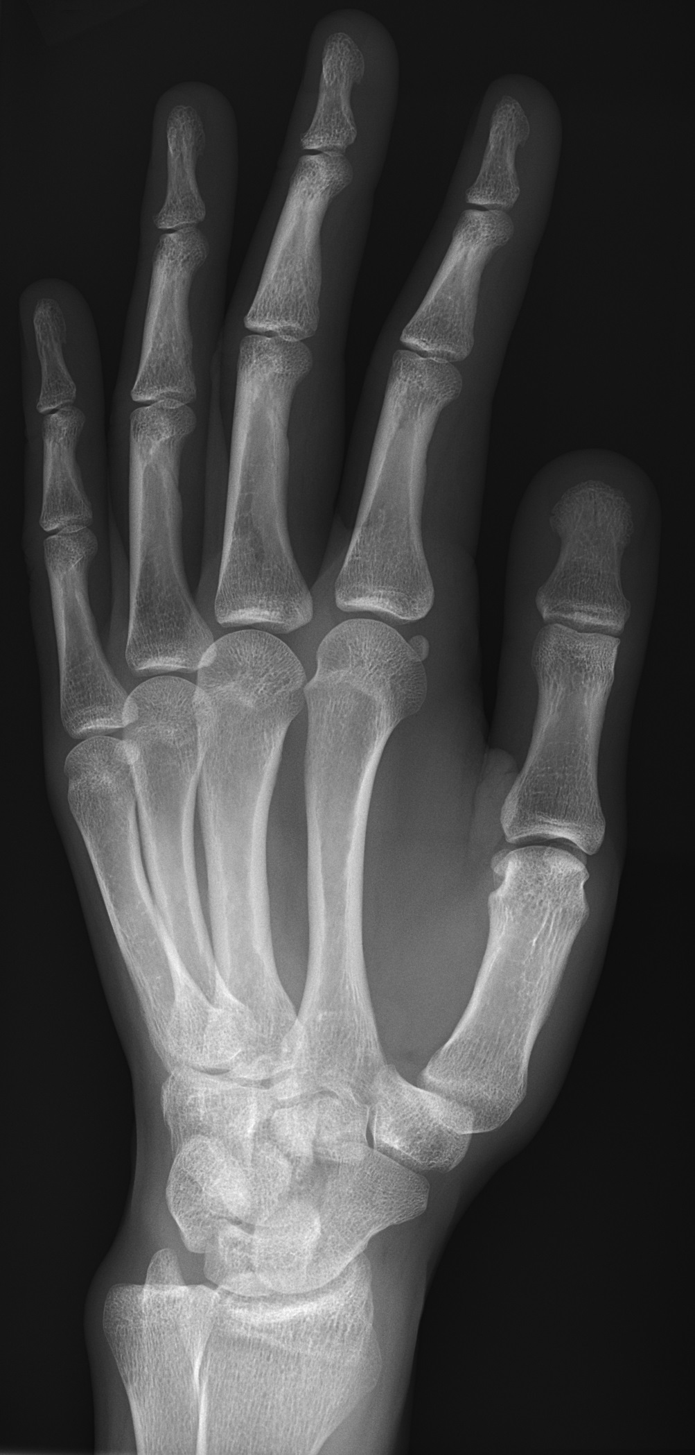
Are currently operating under normal business. MRI is more versatile than the X-Ray and is used to examine a large variety of medical conditions. The bones in the x-ray are compared to the bones of a standard atlas usually Greulich and Pyle.
Fever aches and painsflu-like symptoms. Even with simple sprains the finger may be swollen for up to a full year. Our six locations across the US.
When you get an X-ray taken at a hospital X-ray sensitive film is put on one side of your body and X-rays are shot through you. To produce a chest X-ray test the chest is briefly exposed to radiation from an X-ray machine and an image is produced on a film or into a digital computer. Quite simply PostureRay is a quick and easy radiographic EMR solution specific to todays chiropractor that requires spinal displacements objectively measured and reported on x-ray.
Normal bones pediatric bones normal radiograph normal x-ray normal bones This is a repository of example radiographs x-rays of the pediatric skeleton by age. What is an X-ray of the pelvis. Hover onoff image to showhide findings.
Samples to be analyzed using XRD must be crystalline however the technique can provide the degree of crystallinity in polymers. X-ray shown in Figure 2 of a finger showing a. Example of a slightly rotated not ideal lateral projection of the cervical spine in A and an x-ray of an ideal lateral projection in B.
JSS Medical College Mysuru Normal Chest X-Ray Approach to Chest X-Ray DrVikram Patil Assistant Professor Radiology JSS Medical College and Hospital Mysuru 2. X-ray of the chest also known as a chest radiograph is a commonly used imaging study and is the most frequently performed imaging study in the United StatesIt is almost always the first imaging study ordered to evaluate for pathologies of the thorax although further diagnostic imaging laboratory tests and additional physical examinations may be necessary to help confirm the diagnosis. Tap onoff image to showhide findings.
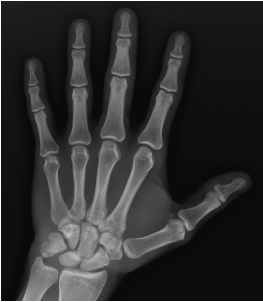
When the condition is caused by trauma it is also known as a boxers knuckle. The red ring shows the position of the External or Lateral epicondyle L which has not yet ossified. Bones of the hand - Normal X-ray PA Hover onoff image to showhide findings.
Tap onoff image to showhide findings. Hand placed palm down in a neutral position with fingers extended on the X-ray plate You can confirm that the handwrist is in a neutral position by drawing a line through the long axis of the radius capitate and the third metacarpal normal axes are within 10 of this line. A hand is easily x-rayed with minimal radiation and shows many bones in a single view.
Magnaflux Hand Held Yokes. CHEST X-RAY TWO VIEWS. Chiropractors have a long history with measuring subluxations by hand on an x-rays utilizing a protractor pencil and specialized plastic templates.
X-ray diffraction XRD is a highly versatile technique that provides chemical information for elemental analysis as well as for phase analysis. The hilar and pulmonary vasculature is normal. They were first discovered and used for imaging purposes by Wilhelm Röntgen on 8th November 1895 when he took an image of his wifes hand.
The Grashey view is obtained with the patient rotated 35-45 degrees so the x-ray beam is parallel to the articular surface of the glenoid. FileRoentgen-x-ray-von-kollikers-handjpg licensed with PD-old. This image is a derivative work of the following images.
It also sees shadows left by things that the X-rays cant travel through like bones or metal. From the American. It occurs when the bones of the finger are moved dislocated from their normal position.
An X-ray or much less commonly X-radiation is a penetrating form of high-energy electromagnetic radiationMost X-rays have a wavelength ranging from 10 picometers to 10 nanometers corresponding to frequencies in the range 30 petahertz to 30 exahertz 30 10 15 Hz to 30 10 18 Hz and energies in the range 124 eV to 124 keVX-ray wavelengths are shorter than those of UV rays and. Bones of the hand - Normal X-ray PA Finger bones articulate at the metacarpophalangeal joints MCPJ the proximal interphalangeal joints PIPJ and the distal interphalangeal joints DIPJ. A more complex method also based on hand x-rays is the TW2 or the TW3 method TW Tanner Whitehouse method.
In water are made to emit a radio signal which are detected by the scanner. Chest X-ray is also referred to as a chest radiograph chest roentgenogram or CXR. X-ray film sees X-rays like the ones that travel through your skin.
The erect anteroposterior chest view is an alternative to the PA view when the patient is too unwell to tolerate standing or leaving the bed 1The AP view examines the lungs bony thoracic cavity mediastinum and great vesselsThis particular projection is often used frequently to aid diagnosis of acute and chronic conditions in intensive care units and wards. Acute traumatic injuries are treated with splinting where chonic injuries often require surgical reconstruction. The potential for ascribing a diagnosis in the setting of a.
Besides chemical characterization XRD is extremely useful for stress measurements as well as for texture analysis. Accessories supplies service and repairs. There is no evidence of any focal area of consolidation.
Click image to align with top of page. A chest X-ray test is a very common non-invasive radiology test that produces an image of the chest and the internal organs. 2008-09-11T194434Z Cropbot 471x738 30859 Bytes upload cropped version operated by UserFinavon.
Original upload log. Finger dislocation is a common injury. The chest radiograph also known as the chest x-ray or CXR is anecdotally thought to be the most frequently-performed radiological investigation globally although no published data is known to corroborate thisUK government statistical data from the NHS in England and Wales shows that the chest radiograph remains consistently the most frequently requested imaging test by GPs 2019 dataset 5.
Body tissues that contain hydrogen atoms eg. Search for magnetic resonance for physics details. Normal elbow X-ray - 10 year old.
Hand-Held Yokes Coils. A dislocated finger can occur in any of the joints of any finger. X-Ray is limited to examining a few body conditions only.
To check for proper alignment look for a normal smooth lordotic curve and imagine two lines each running along the anterior and posterior margins of vertebral bodies. TED carries a huge inventory of Industrial X-Ray equipment and supplies including film film. JSS Medical College Mysuru Introduction Most of the chest x-rays you will see will be normal In order to recognise abnormality you need to know what a normal CXR looks like.
Sagittal band SB rupture leads to leads to dislocation of the extensor tendon of the hand nd may be caused by trauma or by a chronic inflammatory process such as rheumatoid arthritis. All the other centres of ossification are visible.
X Hand Startradiology
1
Normal Hand Radiology Case Radiopaedia Org
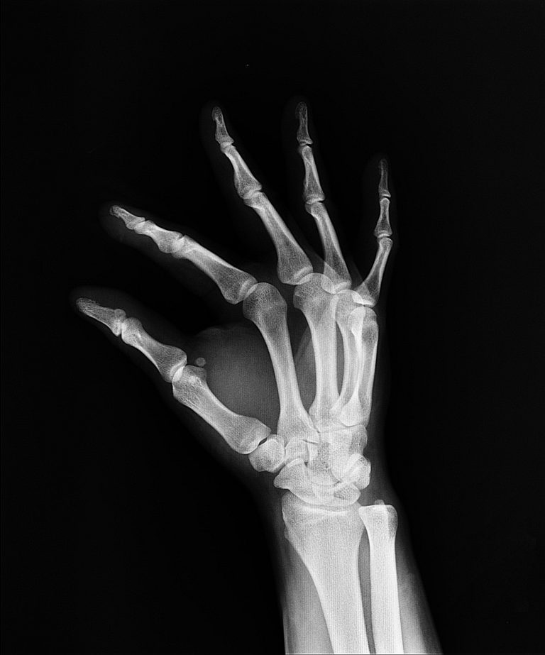
File X Ray Of Normal Hand By Oblique Projection Jpg Wikimedia Commons

File X Ray Of Normal Hand By Lateral Projection Jpg Wikipedia
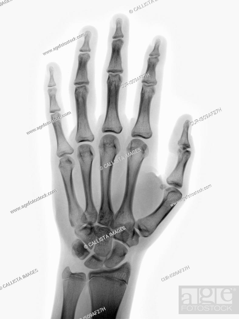
Normal Hand X Ray Of A 15 Year Old Boy Stock Photo Picture And Rights Managed Image Pic Cur Is09af27h Agefotostock
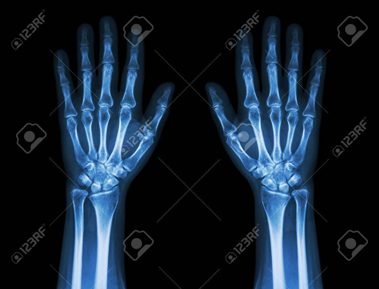
X Ray Hands Front View Normal Human Hands Stock Photo Picture And Royalty Free Image Image 35149286
Xray Image Of Normal Hand Xray Medical Background Stock Photo Download Image Now Istock


